Innovative Studies
As a research scientist in the field of reproductive medicine and genetics, I am grateful for the enriching collaborations that have shaped my professional journey.
I have had the privilege of collaborating with esteemed institutions and organizations worldwide. These collaborations included The Rayne Institute, Richard Dimbleby Laboratory of Cancer Virology, and the Department of Molecular Biology of Fetal Medicine at Queens Charlotte’s Hospital, as well as at the Royal Postgraduate Medical School, Department of Obstetrics and Gynecology, Hammersmith Hospital, all located in London, UK. Additionally, I have had the opportunity to collaborate with Monash Immunology and Stem Cell Laboratories (MISCL) in Melbourne, Australia, and Leeds Institute of Cardiovascular & Metabolic Medicine (LICAMM), UK.
Through these collaborations, we have made groundbreaking advancements that can positively impact the lives of infertile individuals and couples.
I feel honored to have contributed to the growing body of knowledge in this area through significant publications, working alongside leading experts in the field. Each publication I have authored or co-authored reflects my unwavering dedication to expanding scientific knowledge and improving patient outcomes in the field of assisted reproduction.
It is a humbling experience to have the opportunity to contribute to the field and to potentially make a difference in the lives of those on their fertility journey.

With breakthrough studies we’re not just advancing science – we are honoring
our commitment to turning aspirations into cherished realities
Explore my publication timeline
 Published in the Human Reproduction Journal (2005) and titled “Birth of a Healthy Infant following Trophectoderm Biopsy from Blastocysts for PGD of Beta-Thalassaemia Major” this study documented a historic milestone—the pioneering achievement of the first successful birth of a healthy infant using the innovative technique of trophectoderm biopsy from blastocysts for the diagnosis of beta-thalassemia major.
This groundbreaking research laid a robust foundation for the subsequent widespread adoption of blastocyst biopsy as a reliable method for preimplantation genetic testing.
In a world predominantly relying on polar body or cleavage stage biopsy, our study emerged as a pioneering beacon. This achievement not only signaled a remarkable scientific milestone but also set the stage for a monumental paradigm shift. Over the course of approximately five to ten years following our publication, a significant number of medical centers around the globe embarked on a comprehensive review of their established protocols. The consequential outcome of this collective introspection was the widespread adoption of blastocyst biopsy for preimplantation genetic testing—a monumental change that reshaped the landscape of clinical approaches in reproductive medicine.
Our study, thus, encapsulates not only a groundbreaking accomplishment but also the catalyst that propelled the global IVF community toward a new era of enhanced genetic diagnostics and improved patient care.
 Our second study on the pioneering methodology of blastocyst biopsy was published in the Human Reproduction Journal (2007), titled “Blastocyst Biopsy versus Cleavage Stage Biopsy and Blastocyst Transfer for Preimplantation Genetic Diagnosis of Beta-Thalassaemia: A Pilot Study.“ This study was meticulously designed as a randomized control study to compare the efficacy of two methods: blastocyst biopsy and transfer versus cleavage stage biopsy and transfer, within the context of preimplantation genetic diagnosis for beta-thalassemia.
The outcome of our comprehensive investigation notably favored the utilization of blastocyst biopsy and subsequent transfer, revealing superior clinical outcomes. These findings offer significant insights into the potential advantages associated with this approach in the realm of preimplantation genetic diagnosis. Importantly, our study accentuates the potential to improve success rates within assisted reproductive technologies, thus enhancing the prospects of achieving successful pregnancies leading to the birth of healthy offspring.
Upon publication, the impact of our study rippled throughout the scientific community, prompting a significant shift in clinical practice. Over the subsequent years, a substantial number of IVF centers globally undertook a comprehensive reevaluation of their established protocols, with a notable proportion opting to transition from conventional cleavage-stage biopsy to the innovative blastocyst-stage biopsy technique for preimplantation genetic diagnosis. Significantly, our study’s emphasis on the merits of the blastocyst biopsy technique has culminated in its widespread adoption, transforming it into a routine practice across IVF centers worldwide. It not only serves as an informative guide for clinical practice but also propels the trajectory towards optimal methodologies aimed at advancing the outcomes of preimplantation genetic diagnosis
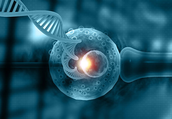
 Published in the Human Reproduction Journal (2008), our research, presented as “A Novel Strategy with the Potential to Identify Developmentally Competent IVF Blastocysts” introduced a groundbreaking methodology to assess blastocyst viability during in vitro fertilization (IVF). This pioneering study aimed to decipher the genetic signatures associated with embryonic development and implantation potential. Through combining blastocyst biopsy, DNA fingerprinting and a comprehensive analysis of gene expression profiles within these blastocysts, our research revealed a compelling link between developmental competence and gene expression patterns pivotal to successful embryo development and implantation.
Such pioneering advancements have garnered significant recognition, with the esteemed Human Reproduction Journal requesting a press release to highlight the importance of our findings. Remarkably, the impact extended well beyond scientific circles, captivating the attention of esteemed media outlets like CNN, Channel 4, Reuters, Bloomberg, and other influential media globally. This research not only introduces a transformative approach, but also opens doors to potential rapid diagnostic assays and enhanced criteria for selecting the most viable blastocyst from a developing in vitro cohort.
In essence, this study’s profound contributions echo widely, offering a promising trajectory for the future of IVF and reproductive medicine. It stands as a pivotal milestone in our scientific journey, igniting progress within the field and reigniting hope for those who hold dreams of nurturing new life into the world.
 Published in the Fetal Diagnosis and Therapy Journal in 2008, this study marks a significant milestone in the field of preimplantation genetic diagnosis. Titled “Birth of a Healthy Histocompatible Sibling following Preimplantation Genetic Diagnosis for Chronic Granulomatous Disease at the Blastocyst Stage Coupled to HLA Typing“ the research aimed to address a compelling family case, involving an affected male child. The primary objective was to identify unaffected embryos that also exhibited HLA compatibility, thereby potentially serving as donors for hematopoietic stem cell transplantation from umbilical cord blood.
Distinguished by its innovative approach, the study achieved unprecedented success. It identified and transferred two healthy, HLA-matched embryos, leading to a singular pregnancy. Remarkably, this success story not only represents a triumph in medical science but also sparked significant societal interest within Greece.
Notably, this pioneering case holds the distinction of being the first instance in Greece to offer preimplantation genetic diagnosis coupled to HLA typing for an affected sibling. This achievement resonated profoundly with Greek society, capturing the attention of both media and press outlets, which extensively covered the breakthrough. In essence, this research not only underscores the transformative potential of advanced preimplantation genetic testing methodologies but also highlights the power of scientific progress in instilling hope among families grappling with complex genetic conditions.
 Published in the Biology of Blood and Marrow Transplantation Journal (2010), our work titled “Successful Hematopoietic Stem Cell Transplantation in Children with X-linked Chronic Granulomatous Disease from HLA-Identical Siblings Selected through Preimplantation Genetic Diagnosis Combined with HLA Typing“ represents a significant stride in medical achievement. We share, before many others that followed in subsequent years, the remarkable cases of two children afflicted with X-linked chronic granulomatous disease who underwent successful hematopoietic stem cell transplantation using grafts from their unaffected and HLA-matched siblings, thoughtfully chosen before implantation.
Using preimplantation genetic diagnosis in conjunction with HLA-matching, specific embryos were selected for intrauterine transfer. As a result, two singleton pregnancies developed to term, yielding a healthy female and carrier female, both sharing HLA-identical traits with their affected siblings in their respective families. The recipient siblings, aged 4.5 years and 4 years, underwent combined umbilical cord blood and bone marrow stem cell transplantation, yielding promising results of complete donor hematopoietic and immunologic reconstitution during follow-up assessments.
This groundbreaking accomplishment marked a pioneering moment for Greece, leaving an indelible impact on Greek society. The widespread media coverage of our research deeply resonated with countless couples who find themselves confronting similar medical challenges.
Our work not only represents a significant scientific leap but also serves as an uplifting testament to research’s capacity to positively shape society. By providing tailored medical solutions to families grappling with genetic disorders, we enhance the lives of those affected while contributing to a deeper comprehension of genetic conditions.
As I contemplate the very essence of our work, I am deeply touched by a profound message of hope and inspiration. When I recall the stories of the mothers, I have had the privilege of meeting over the years, they stand as a powerful testament to the unwavering strength of maternal love in their determined journey toward healing and wholeness. This serves as a gentle yet powerful reminder of the transformative magic that weaves through the fabric of science, love, and the unbreakable bonds that embrace a family.
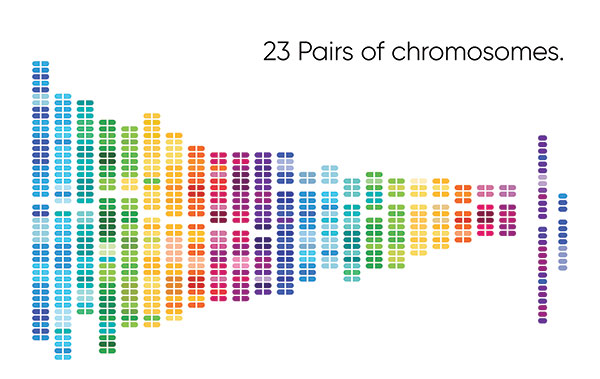 Published in Human Reproduction in 2011, the study titled “PGD for Reciprocal and Robertsonian Translocations using Array Comparative Genomic Hybridization“ introduced the clinical application of array comparative genomic hybridization (array-CGH) as a molecular-based technique for detecting unbalanced chromosome rearrangements and aneuploidy in preimplantation embryos. Array-CGH had not been used for preimplantation genetic testing of structural rearrangements at that time, and its needed to be validated.
The Study demonstrated that, in contrast to the widely employed traditional FISH-based methods at that time, array-CGH not only exhibited enhanced test performance, sensitivity, and reliability but also provided the notable advantage of simultaneous screening for aneuploidy across all 24 chromosomes.
This groundbreaking research represented a significant leap forward in the realm of preimplantation genetic testing, serving as one of the early publications that underscored the potential of Array-CGH. Subsequently, numerous researchers have delved into and contributed to the advancement of this dynamic area of study.
 I had the privilege of being interviewed about current practices and perspectives related to preimplantation genetic screening (PGS), with a specific focus on its newest form: PGS 2.0. This occurred as part of a significant research initiative titled “The Why, the How, and the When of PGS 2.0“ which was subsequently published in the Molecular Human Reproduction journal in 2016. The primary aim of this study was to thoroughly investigate the diverse range of opinions and opinions and current practices on preimplantation genetic screening. The selection process for experts were based on stringent criteria, which encompassed their proven expertise in PGS as substantiated by peer-reviewed publications and active participation in prestigious scientific forums such as the European Society of Human Reproduction and Embryology and the American Society for Reproductive Medicine meetings.
As an embryologist expert, my involvement in this study centered on the aspect of “The When.” My insights and perspectives were sought to contribute to a comprehensive understanding of the optimal timing for the implementation of PGS 2.0.
The study stands as an invaluable repository of insights into the evolving PGS landscape. It offers a snapshot of prevalent practices and perspectives during the time of its publication, while also acknowledging the ongoing discourse within this dynamic field. Importantly, the insights gleaned from the study have positioned it as a valuable reference for fertility specialists and researchers alike, providing a comprehensive overview of the diverse perspectives surrounding PGS 2.0 and its multifaceted implications.
 I had the distinct honor of being appointed as the Research Coordinator for the Athens Group, one of only eight prestigious centers worldwide selected to participate in the groundbreaking study titled “Preimplantation Genetic Testing for Aneuploidy by Microarray Analysis of Polar Bodies in Advanced Maternal Age: A Randomized Clinical Trial.“ This pivotal research, published in Human Reproduction Journal in 2018, truly embodies our commitment to advancing the frontiers of reproductive medicine.
Spanning the years from 2012 to 2016, the ESTEEM trial marked a significant milestone as a multinational, multicenter, pragmatic, randomized clinical trial, employing intention-to-treat analysis involving patients recruited from nine centers in seven countries across Europe and Israel. These centers, recognized for their proven expertise in polar body biopsy and array comparative genomic hybridization analysis, collectively embarked on the mission to estimate the efficacy of polar body microarray analysis for preimplantation genetic testing in women aged 36-40 undergoing ICSI treatment, in broad routine clinical practice.
While the study did not show a substantial increase in the live birth rate for the chromosome screening group, it did reveal potential benefits, such as fewer transfers, reduced cryopreservation, and decreased miscarriages.
Our involvement in this study underscores our commitment to pioneering research, aimed at informing, and enhancing fertility treatments. Through our dedicated engagement, we contribute to a deeper understanding of the benefits and implications of preimplantation genetic testing for aneuploidy. Ultimately, our goal is to empower women navigating age-related fertility challenges, equipping them with the knowledge and options they need to make well-informed decisions on their journey towards parenthood.
 I take great pride in our research endeavor published in 2019, titled “Can Trophectoderm RNA analysis predict human blastocyst competency?“ This work was featured in the esteemed journal “Systems Biology in Reproductive Medicine.”
Building upon our 2008 research that examined blastocyst expression profiles linked to implantation, we embraced a new direction in 2019. Instead of traditional microarrays, we adopted an ultra-sensitive single-cell RNA sequencing method. Our core aim was to determine whether highly sensitive RNA-seq analysis of blastocyst biopsies could effectively distinguish between blastocysts capable of successful implantation and those that were not.
By implementing this advanced RNA sequencing strategy, our goal was to identify a more robust set of markers capable of accurately assessing blastocyst implantation competence. Our pilot study, combined with complementary research findings, indicated the potential of trophectoderm (TE) biopsy in predicting blastocyst competency. This method should undergo comprehensive evaluation for its applicability in this specific context.
It is worth noting that this work demanded an additional two years of effort before we could proceed with the patent application. For further details on developments in 2021, please refer to the corresponding section.
 I was deeply honored to receive a prestigious invitation from ESHRE to lead the working group for the development of good practice recommendation for polar body and embryo biopsy in preimplantation genetic testing. The scientific paper published in Human Reproduction Open in 2020, titled “ESHRE PGT Consortium and SIG Embryology Good Practice Recommendations for Polar Body and Embryo Biopsy for PGT†.“
Given that the prior ESHRE guidelines for Preimplantation Genetic Diagnosis had become outdated, our mission was to provide up-to-date and comprehensive recommendations that encapsulated the latest advancements and insights within the field. As part of a series of papers on PGT best practices, this publication focused specifically on technical aspects of embryo biopsy, covering the entire spectrum from procedural details to technical recommendations related to biopsy, cryopreservation, and laboratory practices.
Our effort not only contributed to the advancement of knowledge within the scientific and medical community but also encapsulated our commitment to shaping the future of preimplantation genetic testing through meticulous research and shared expertise.
The review report and the corresponding webinar are accessible on the ESHRE website.
Please follow the link provided below.
https://www.eshre.eu/Guidelines-and-Legal/Guidelines/PGT
https://ecampus.eshre.eu/pluginfile.php/43755/mod_kalturavideo/content/0/webinar16062020.pdf
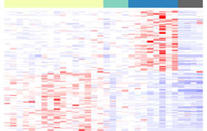 I take immense pride in our research project titled “The Impact of Aging on Molecular Modulators of Human Embryo Implantation“ published in Science journal in 2021. This pride stems from the fact that our findings have ignited a beacon of hope, laying the path for a diagnostic test that could bring dreams to life for women undergoing fertility treatment.
This pioneering research successfully unveiled biomarkers that are consistent with embryo-endometrial crosstalk/developmental competency and serve as mediators for successful implantation. Furthermore, we uncovered potential roles for extracellular exosomes, embryonic metabolism, and the regulation of apoptosis.
Significantly, our study represented a high-throughput approach to examine embryo-endometrial interactions without necessitating unethical in vivo experimentation on humans. The experimental design employed here offered a window into how aging orchestrates its changes in the dynamic realm of the trophectoderm transcriptome.
The implications of our work are far-reaching. Our identification of these biomarkers could potentially serve as the foundation for a clinical diagnostic test. This test would enable the selection of the most competent blastocysts for uterine transfer, enhancing the chances of successful implantation and subsequent development. This achievement led us to initiate a patent application (application number: GB 2107813.4), a significant milestone that brings both gratification and responsibility to our entire team.
Stay tuned for more exciting developments in this captivating field!
 Published in the Human Reproduction Journal in 2020, our study entitled “Age-Related Transcriptomic Changes During the Germinal Vesicle to Metaphase II Transition in Euploid Human Oocytes“ marks a significant milestone in our ongoing exploration of reproductive science. This investigation delves into gene expression shifts within euploid human oocytes, particularly as they undergo the pivotal transition from the germinal vesicle (GV) to the metaphase II (MII) stage. Our research takes a focused look at the influence of age on these changes, bringing valuable insights to individuals with advanced age.
An intriguing revelation emerged from our findings: a discernible decrease in mitochondrial-associated transcripts during this crucial transition. What caught our attention even more was the pronounced impact on MII oocytes from individuals with advanced age. As we recognize the paramount role of maternal transcripts in the early stages of embryonic development, our study contributes to the understanding of why oocytes from advanced maternal age may exhibit reduced competence. This potentially stems from age-related shifts in transcriptional activity.
This research represents a significant step forward, unraveling the intricate relationship between aging and the transcriptome of euploid oocytes. The implications extend to fertilization and early embryo development. To solidify our insights, we employed a variety of methodological approaches, which hold the promise of identifying molecular markers to assess oocyte quality.
Stay tuned as we navigate deeper into the realm of reproductive science for more updates and insights!
 Building upon our prior research, our latest work titled “Trophectoderm Non-Coding RNAs Reflect the Enhanced Metabolic and Invasive Characteristics of Blastocysts from Young Maternal Ages“ is published in the 2023 edition of Systems Biology in Reproductive Medicine.
Our investigation stems from the understanding that advancing female age coincides with reduced fertility, due to genetic, environmental, and dietary factors across a lifetime. Our investigation delves into both coding and non-coding RNAs in embryos, aimed at a deeper comprehension of the root causes and implications of the physiological changes accompanying this decline.
Summarizing our discoveries, the identified non-coding RNA biomarkers align with processes involving embryo-endometrial crosstalk, developmental competence, and implantation. The corresponding genes associated with expressed non-coding RNAs display heightened activity in blastocysts from younger women and are associated with metabolic pathways, particularly cholesterol biosynthesis and steroidogenesis.
This work brings greater clarity to the intricate connections between maternal age, blastocyst properties, and the underlying molecular processes, further enriching our understanding of reproductive dynamics.
Stay tuned for upcoming insights!

